Researchers have developed a simple, non-invasive method of diagnosing Parkinson’s disease early using an eye scan. It could mean treatment aimed at slowing the disease can start sooner or, alternatively, could be used to monitor the disease’s progression.
As is the case with many progressive conditions, the earlier that Parkinson’s disease is diagnosed, the better. Early diagnosis means that interventions can be introduced ahead of time that may prevent or curtail the disease’s destructive course. Unfortunately, most Parkinson’s diagnoses are made after the emergence of symptoms.
Researchers from Université Laval and its affiliate, the CERVO Brain Research Center, both located in Québec, Canada, have developed a method of diagnosing Parkinson’s disease (PD) early, using a simple and non-invasive eye scan.
“By [the time symptoms appear], the disease has been present for several years and the affected neurons are already engaged in an irreversible degenerative process,” said the study’s corresponding author, Martin Lévesque, PhD, professor at Laval’s Faculty of Medicine and CERVO researcher. “That’s why it’s important to find biomarkers that detect Parkinson’s at an early stage of the disease. An unusual response of the retina to light stimuli could be indicative of a pathology affecting the brain.”
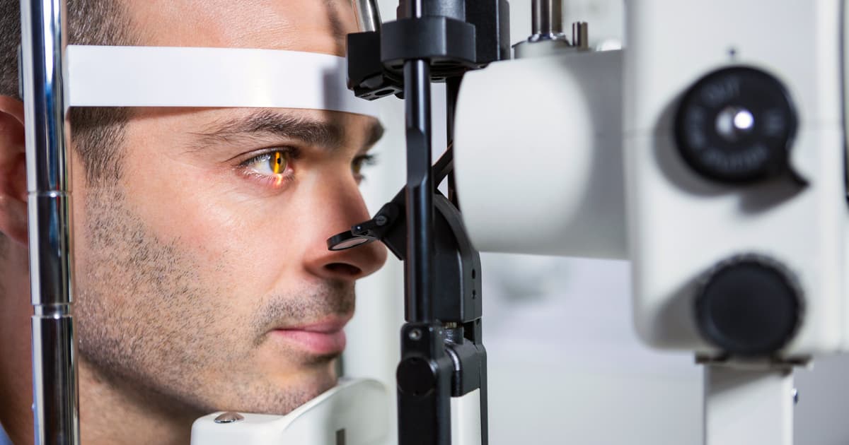
In recent years, visual impairments arising from retinal dysfunction have gained attention as potential indicators of PD. Electroretinography (ERG) is a technique used to evaluate the electrical activity of the retina, the light-sensitive tissue at the back of the eye, in response to light stimulation. A device delivers light flashes or patterns to the eye, and electrodes placed on and around the eye measure the electrical responses generated by different types of retinal cells to give insight into how well they are functioning.
While ERG has been used to reveal subtle retinal changes that correlate with psychiatric disorders such as schizophrenia, bipolar disorder, and depression, its use has not been widely researched in the setting of PD. In the present study, the researchers hypothesized that distinct ERG impairments could serve as early Parkinson’s indicators. They tested their hypothesis on 20 adults who had been diagnosed with PD within the last five years, and 20 age-matched adults without PD.
“We placed an electrode on each participant’s lower eyelid and recorded their retinal response to a series of flashes of different intensity, frequency and color,” Lévesque said. “We did the same with people of the same age, but in good health. The results we obtained for people with Parkinson’s had a distinct signature from those in the control group.”
The researchers also used ERG to test their theory on two-month-old M83 transgenic mice, genetically engineered to produce a mutated form of the human alpha-synuclein protein associated with familial forms of PD. M83 mice develop motor impairments and neurological symptoms like those seen in human patients with Parkinson’s. The results of ERG testing on these mice were compared with those of an age-matched control group.
“We used young mice in which no motor signs of the disease were yet observable,” said Lévesque. “Once again, we obtained different responses in Parkinson’s model animals. This suggests that the functional manifestations of Parkinson’s could be detected at an early stage of the disease by retinal examination.”
The average age of Parkinson’s diagnosis is 65. The researchers hope that this simple and non-invasive test can be used to start diagnosing PD earlier. Alternatively, it can be used to keep an eye on the disease’s progression.
“We could offer a functional retinal exam from the age of 50,” Lévesque said. “By detecting the disease early, we could offer interventions that prevent the degeneration of the neurons involved in Parkinson’s. This approach could also be used to monitor the progression of the disease, as well as the effectiveness of interventions offered to patients.”
The study was published in the journal Neurobiology of Disease.
Source: Université Laval, EurekAlert!
Get the source article here