This approach could also be used to monitor the progression of the disease, as well as the effectiveness of interventions offered to patients
Martin Lévesque
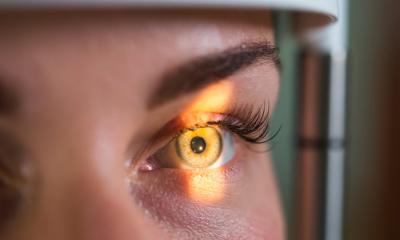
To explore this avenue, his research team recruited 20 people who had been diagnosed with Parkinson’s for less than 5 years. “We placed an electrode on each participant’s lower eyelid and recorded their retinal response to a series of flashes of different intensity, frequency and color. We did the same with people of the same age, but in good health. The results we obtained for people with Parkinson’s had a distinct signature from those in the control group,” explains Professor Lévesque.
The research team carried out similar tests in transgenic mice overexpressing a human protein associated with Parkinson’s disease. “We used young mice in which no motor signs of the disease were yet observable. Once again, we obtained different responses in Parkinson’s model animals. This suggests that the functional manifestations of Parkinson’s could be detected at an early stage of the disease by retinal examination”, sums up Martin Lévesque.
Parkinson’s disease occurs most often in people over the age of 60. “We could offer a functional retinal exam from the age of 50,” continues Lévesque. “By detecting the disease early, we could offer interventions that prevent the degeneration of the neurons involved in Parkinson’s. This approach could also be used to monitor the progression of the disease, as well as the effectiveness of interventions offered to patients.”
The study’s first author is doctoral student Victoria Soto Linan. The coauthors are Véronique Rioux, Modesto Peralta III, Nicolas Dupré, Marc Hébert and Martin Lévesque.
Source: Université Laval